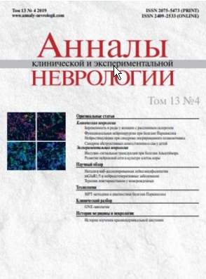
Vol 13, No 4 (2019)
- Year: 2019
- Published: 26.12.2019
- Articles: 12
- URL: https://www.annaly-nevrologii.com/journal/pathID/issue/view/63
Full Issue
Original articles
Analysis of pregnancy and childbirth in women with multiple sclerosis: a prospective study
Abstract
Introduction. Family planning for patients with multiple sclerosis (MS) raises many questions and requires an integrated approach from neurologists and obstetrician-gynaecologists.
The study aimed to define possible features of pregnancy and childbirth in patients with MS.
Materials and methods. 204 patients with definite moderate MS, who were planning a pregnancy and taking First-Line Disease-Modifying Therapies (DMTs) before pregnancy. First group included 94 patients with pregnancies; the second group consisted of 110 patients, who failed to conceive within a stated period of time; in the third group there were 50 healthy women with normal pregnancies. Probability of developing pregnancy complications, time and method of delivery, anaesthetic procedures, weight, and height of newborns were assessed, while in the groups of patients with MS the risk of exacerbations and severity of the complications were evaluated.
Results and discussion. In the first group, there were more frequent threats of miscarriage and preterm birth, which might have taken place due to a complex of factors (cancellation of DMTs, use of hormonal therapy for exacerbations during pregnancy). The high frequency of caesarean section in the first group was associated with the unreasonable alertness of obstetrician-gynaecologists and the fear of the patients that the course of MS would worsen. Frequent exacerbations during pregnancy were associated with the abolition of DMTs before pregnancy and the failure of physiological immunosuppression during pregnancy.
Conclusion. In Russia, there is no single protocol for managing patients with MS during the period of family planning, pregnancy, and the postpartum period, which causes certain difficulties. There is emerging evidence that certain DMTs can be prescribed during pregnancy and lactation, which will help minimize the risks of exacerbations and disability increase.
 5-9
5-9


Spinal cord stimulation in failed back surgery syndrome
Abstract
Introduction. A common cause of chronic pain after back surgery is failed back surgery syndrome, characterized by the development, persistence or recurrence of neuropathic pain in the absence of clear anatomical complications from the surgery. One of the most effective treatment methods for failed back surgery syndrome is permanent spinal cord stimulation.
Study aim. To assess the efficacy and safety of chronic spinal cord stimulation in failed back surgery syndrome.
Materials and methods. In our study, after a stimulation trial final neurostimulation was performed in 34 patients with neuropathic pain and lack of improvement from the pharmacological treatment.
Results. Six months after the operation, there was a reduction in the average scores on the visual analogue scale for the daily average and maximum pain, as well as a reduction in the severity of neuropathic pain as measured by the PainDetect scale (by 54.4% 50.7% and 57.3%, respectively), which corresponds to the method’s efficacy criteria found in the literature. The majority of patients noted a significant improvement in their quality of life and a reduced need for pain relief. Complications occurred in 26.4% of patients overall, including intraoperative damage to the dura mater, infection at the generator implantation site, and electrode displacement relative to the initial position, requiring correction. None of the patients experienced worsening of the neurological symptoms.
 16-22
16-22


Molecular expression of insulin signal transduction components in brain cells in an experimental model of Alzheimer’s disease
Abstract
Introduction. The risk of Alzheimer’s disease (AD) is increased with cerebral insulin resistance, which may be caused by the impaired function of the cerebrovascular system, and may also have a direct effect on β-amyloid aggregation and Tau protein phosphorylation.
Aim. To study the molecular expression of insulin signal transduction components (IRS1, GSK3B and PKC) in the brain cells in an experimental model of AD.
Materials and methods. Experiments were conducted on 4-month-old C57BL/6 and B6.129S6-Nlrp3tm1Bhk/JJ male mice (NLRP3 knockout mice) with 5 animals in each group. AD was modelled in the experimental group of mice by administering β-amyloid; mice in the control group received sham surgery. IRS1, GSK3B and PKC expression in the amygdala was studied using immunohistochemistry methods.
Results. The C57BL/6 mice with AD had reduced IRS1 expression compared with the mice who received sham surgery (0.62±0.13 and 0.89±0.17; р=0.045), while the β-amyloid did not produce the same result in NLRP3 knockout mice. GSK3B expression was increased in C57BL/6 mice with AD (0.60±0.12) when compared with both the control group (0.20±0.02; p<0.0001) and the NLRP3 knockout mice with AD (0.27±0.08; p<0.0001). PKC expression in C57BL/6 mice with AD was reduced (0.52±0.14) when compared with the NLRP3 knockout mice with AD (0.89±0.18; p<0.05) and the control group (0.84±0.12; p<0.05).
Conclusion. The development of Alzheimer type-neurodegeneration is accompanied by disruptions in IRS1 and GSK3B expression, which is associated with impaired signal transmission along the PKC pathway. The suppression of neuroinflammation through NLRP3 inflammasome deletion has a protective effect in AD.
 28-37
28-37


Changes in the morphofunctional development of the neuronal network in a dissociated cell culture of rat cerebral cortical neurons
Abstract
Introduction. Study of the morphofunctional neuronal development in a dissociated cerebrocortical cell culture, using modern cell technologies, is a priority in experimental neurology, which is required for successful in vitro modelling of acute and chronic forms of cerebral pathology.
Aim. A morphofunctional study of the in vitro changes in neuronal differentiation of rat cerebral cortical neurons, using a range of analysis methods, including immunohistochemistry, fluorescence, and electrophysiology.
Materials and methods. We investigated the degree of culture differentiation on day 3–4 and day 10–11 of in vitro cultivation, measured by the intensity of the PSA-NCAM protein expression and the level of neuronal glutamate-induced calcium overload. That was then compared with the functional activity of the neuronal network cultivated on a microelectrode array, and with changes of the neuronal network’s activity in response to glutamate receptor overstimulation.
Results. A significant glutamate-induced increase of the intracellular calcium concentration was typical for mature neurons (day 10–11 of cultivation), along with a lack of PSA-NCAM paranuclear accumulation, which was only found in immature cells (day 3–4 of cultivation). There was a glutamate suppression of the neuronal network burst activity, formed in vitro by day 10–11, with had no effect on the generation of single action potentials. At the same time, kainate, the exogenous selective agonist of the one of the glutamate subtypes, completely blocked spontaneous activity of the mature neurons.
Conclusion. Neocortical rat neurons reach the differentiation level necessary for the modelling of the cerebral pathologies by day 10–11 of in vitro cultivation. At this point, the process of disruption of the microelectrode array cultivated neuronal network by the glutamate receptor overactivation, has become multilayered: excitotoxic glutamate-induced damage produces selective disruption of neuronal burst activity, and with the greater cytotoxicity caused by kainate, spontaneous bioelectrical activity is completely blocked.
 38-45
38-45


Reviews
Functional neurosurgery in Parkinson’s disease in Russia
Abstract
The article presents an overview of the most topical matters relating to functional neurosurgery in Parkinson’s disease: the historical aspects, an overview of the international recommendations, long-term effects of neurostimulation, and the selection criteria for the surgical intervention. Summary data are provided for deep brain stimulation surgery in Russia, as well as an analysis of the need for neurostimulation compared with the international data.
 10-15
10-15


Clinical features and mechanisms of the development of cognitive impairment in children with obstructive sleep apnoea syndrome
Abstract
The article reviews clinical features and mechanisms of the development of cognitive impairment in children with obstructive sleep apnoea/hypopnea syndrome (OSAHS). Short-term and long-term consequences of sleep apnoea along with the pathogenetic similarity of OSAHS and neurodynamic disturbances and concomitant conditions are discussed. The role of ENT conditions in children with OSAHS is reviewed as well. Data on the genetic features that affect the risk of developing OSAHS in children are presented.
 23-27
23-27


Natalizumab-associated progressive multifocal leukoencephalopathy in patients with multiple sclerosis: risk reduction, management and possibilities for subsequent immunoregulation
Abstract
The risk of progressive multifocal leukoencephalopathy (PML) is one of the factors limiting the widespread use of natalizumab (NTZ). As a strategy for potentially reducing the frequency of the NTZ-associated PML, while maintaining the high therapy efficacy, in addition to the widely-used risk stratification strategy based on anti-JC virus antibody index and therapy duration, we propose the administration of NTZ with an extended dosing interval. Opinions vary on the effectiveness of plasmapharesis in confirmed cases of PML. The most challenging problem is how to manage patients, who develop PML, and what MS disease-modifying therapy should be considered for the subsequent use.
 46-53
46-53


Group I metabotropic glutamate receptors (mGluR1/5) and neurodegenerative diseases
Abstract
This overview describes how group mGluR1/5 metabotropic glutamate receptors are involved in neurodegenerative diseases; it also touches upon their use as therapeutic targets in animal models. mGluR1/5 are primarily located on the neuronal postsynaptic membrane, where they communicate with two proteins, Gαq/11 and Homer, which, in turn, initiate several biochemical cascades. The Gαq/11 protein cascade includes Са2+ release from the endoplasmic reticulum (ER) through the inositol trisphosphate receptors (IP3R) and the activation of depot-controlled Са2+ entry. The Gαq/11 protein cascade also includes the production of diacylglycerol with subsequent activation of various protein kinases, which, in turn, provide influences on the genome. The Homer protein communicates directly with the NMDA receptors and Shank scaffold proteins, through which it regulates the activity of various protein kinases, including Akt and ERK1/2. The activation of mGluR1/5 triggers long-term depression of glutamatergic transmission through the endocytosis of AMPA receptors, caused by changes in the level of protein phosphorylation and genome activation.
It is thought that mGluR1/5 play an important role in the development of neurodegenerative diseases. In Alzheimer's disease, mGluR1/5 acts as a target for the β-amyloid peptide. mGluR1/5 antagonists have a neuroprotective effect in transgenic mice with Alzheimer's disease. The pathogenesis of Alzheimer's disease includes increased Са2+ release from the ER due to the pathological activity of mGluR1/5, as well as the influence of mutated presenilin on Са2+ homeostasis in the ER. At the same time, restoration of Са2+ levels in the ER is disrupted by the effect of presenilin on depot-activated Са2+ entry.
mGluR5 (but not mGluR1) is being studied as a potential therapeutic target in Parkinson's disease. Numerous studies on rodent and primate models of Parkinson's disease have demonstrated a significant antiparkinsonian effect when mGluR5 antagonists were used. It is thought that the neuroprotective mechanisms of action of mGluR5 antagonists involve limiting the increase in intracellular Са2+ by reducing IP3 and NMDA receptor activation. Huntington’s disease is related to a mutation in the HTT gene and the ability of the mutant huntingtin protein to sensitise IP3 and NMDA receptors, thus triggering Са2+ overload in the neurons. A neuroprotective effect in transgenic mice with Huntington’s disease was achieved by using positive allosteric modulators of mGluR5, capable of selectively activating cascades associated with the Homer protein and triggering Akt activation.
 54-64
54-64


Therapeutic drug monitoring of levetiracetam in newborns
Abstract
Neonatal seizures in full-term and preterm infants represent a common neurological syndrome. Levetiracetam (LEV) is one of the new and widely prescribed second- or third-line antiepileptic drug for the treatment of seizures. Routine therapeutic drug monitoring of LEV was not recommended due to its almost ideal pharmacokinetic profile: linear pharmacokinetics, predictable dose-concentration relationship, wide therapeutic index, favourable safety profile, and unlikely clinically significant drug-drug pharmacokinetic interactions.
In newborns, drug pharmacokinetics may be under the influence of maturation process. LEV pharmacokinetics in newborns appears to be age (gestational, postnatal) dependent and highly variable within the age ranges. These aspects make therapeutic drug monitoring a useful procedure for therapy optimization in this specific patient population.
In population modeling based on therapeutic drug monitoring and nonlinear mixed effects models, covariates were found that should significantly affect the LEV clearance and volume of distribution in newborns — creatinine clearance and total body weight. Using of these regression equations can help to adjust the LEV doses without the patient's measured concentration data. But the significant magnitudes of the interindividual variability remaining in these final regression models justify the need for therapeutic drug monitoring and Bayesian adaptive control for personalization of LEV dosage regimens in neonates.
 65-76
65-76


Technologies
New MRI diagnostic methods in Parkinson's disease: evaluating nigral degeneration
Abstract
Research Center of Neurology, Moscow, Russia
Parkinson's disease (PD) is a progressive neurodegenerative disorder with a characteristic pathological hallmark of loss of the dopaminergic neurons in the compact part of the substantia nigra in the midbrain. Despite the significant progress made in learning about this disease, early diagnosis continues to be a complex clinical issue. Currently, many studies are focused on finding and implementing meaningful markers which are valid for the early PD diagnosis. One of the most promising areas in that field is investigation of specific changes in the substantia nigra, found when examining the nigrosomes (specific clusters of the dopaminergic neurons) and neuromelanin, using high-field magnetic resonance imaging (MRI).
This article presents the current understanding of the structural and functional organization of the substantia nigra, and examines in detail the new informative MRI-markers of neurodegeneration in PD: the loss of dorsolateral nigral hyperintensity (disappearance of the nigrosome-1) and a reduction in the intensity/area of the magnetic resonance signal from the substantia nigra when imaging the neuromelanin. We present our own experience of using the abovementioned technologies to diagnose PD, by analysing susceptibility-weighted images and images taken in neuromelanin-sensitive MRI mode.
 77-84
77-84


Clinical analysis
GNE myopathy (Nonaka myopathy)
Abstract
GNE myopathy (Nonaka myopathy) is a rare recessive muscular dystrophy associated with the GNE gene, which is involved in sialic acid synthesis. Typical onset is in the third decade of life with distal weakness of the arms and legs, gradually progressing to the proximal muscles, along with severe generalized myopathy and loss of ambulation usually occurring 10–20 years after disease onset. Exomе sequencing methods have greatly increased the possibility of diagnosis of this and other rare hereditary diseases. A case of GNE myopathy with onset at 26 years of age and a prolonged search for a diagnosis, which was finally made after 12 years, is presented. Whole exome sequencing with subsequent Sanger sequencing verification found compound heterozygosity of the GNE mutations not previously described: с.787delA (p.Met263CysfsTer4) and c.2005G>T (p.Ala669Ser). Differential diagnosis and a literature review are presented.
 85-90
85-90


Historical articles
Historical aspects of studying craniocervical dystonia
Abstract
The study of dystonic hyperkinesias has a thousand-year-old history. Beginning with drawings and sculptures from antiquity and up to the present day, modern ideas gradually developed about the phenomenology, origin, and treatment methods of dystonia. Mentions of spastic torticollis, blepharospasm and Meige syndrome can even be found in the writings of Hippocrates and Celsus. Images and monuments from antiquity and ancient civilizations indicate the existence of focal dystonias in those times.
The Middle Ages left science with records of cervical dystonia and numerous illustrations in religious images. The first well-known mention of the term ‘torticollis’ belongs to François Rabelais. The term started to appear in medical texts later on. One of the earliest medical records on cervical dystonia was made by the Swiss physician Felix Platerus. During the Age of Enlightenment, dystonias became a separate class in disease classification.
Modern tendencies in studying dystonia are characterized by identifying the genes responsible for different forms of primary dystonia along with description of their phenotypes. There is an ongoing research on the role of mental disorders in the clinical presentation of dystonia.
 91-96
91-96


