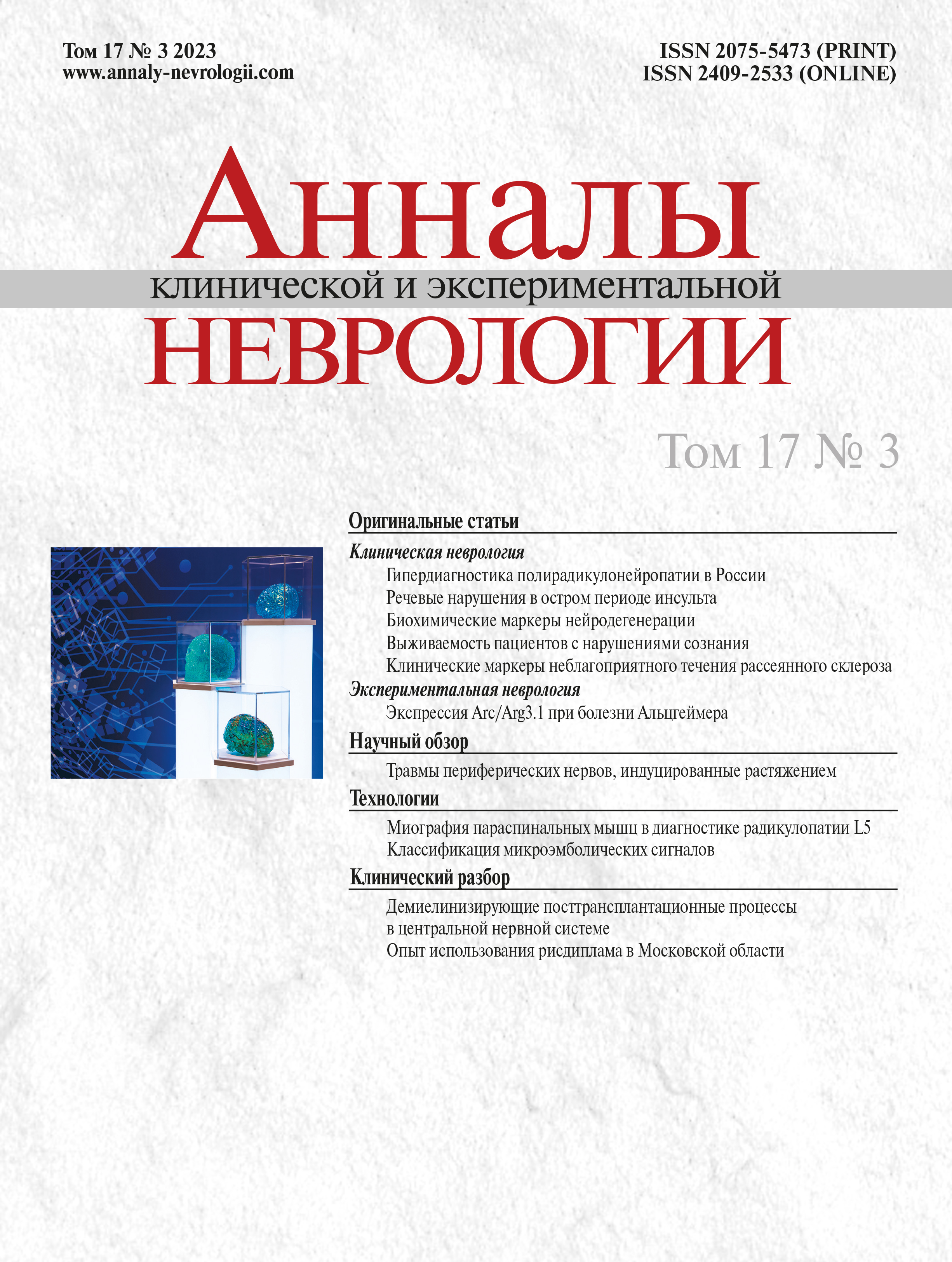
Vol 17, No 3 (2023)
- Year: 2023
- Published: 29.09.2023
- Articles: 12
- URL: https://www.annaly-nevrologii.com/journal/pathID/issue/view/79
Full Issue
Original articles
Chronic inflammatory demyelinating polyradiculoneuropathy overdiagnosis in Russia
Abstract
Introduction. Despite the improving diagnostic criteria for the chronic inflammatory demyelinating polyradiculoneuropathy (CIDP), its verification is still an issue.
Objective: to study the rate and the causes of CIDP misdiagnosis.
Materials and methods. We prospectively and retrospectively analyzed the clinical and paraclinical data of 223 patients admitted to the Research Center of Neurology from 2018 to 2022 with a CIDP referral.
Results. We revised the CIDP diagnosis in 150/223 patients (67%; median age 55.5 [43; 63] years; 75 males and 75 females; 3-year follow-up history [1.75; 5.25].) Once the definitive diagnosis was clarified, we divided the patients into the following groups: polyneuropathy of other etiology (n = 94; 63%), other neuromuscular disorders (n = 39; 27%), CNS disorders (n = 10; 7%), no structural NS disease (n = 7; 5%). Patients did not meet the 2021 EAN/PNS diagnostic criteria at the history-taking stage in 65% of cases, at the neurological examination stage in 39% of cases, and at the electroneuromyography stage in 92% of cases.
Conclusions. The rate of CIDP misdiagnosis in Russia is 67%, and most often this refers to patients with polyneuropathy of other etiologies. The main cause for the CIDP misdiagnosis was inaccurate electroneuromyography. We should bear in mind that CIDP is a rare disorder with an extensive differential diagnosis, so it should be verified according to the current 2021 EAN/PNS diagnostic criteria.
 5-12
5-12


Features of speech disorders in patients with acute ischemic stroke
Abstract
Introduction. Various speech disorders that lead to impaired communication occur in 30–50% of ischemic stroke (IS) survivors. Although most attention is traditionally paid to aphasia, speech disorders also include the following: dysarthria, dysphonia (isolated or in combination with dysarthria and/or dysphagia), fluency disorders, and non-specific speech disorders associated with the severity of condition and a cognitive disorder.
Objective: to study the variety of speech disorders and their features in patients with acute IS.
Materials and methods. We examined 69 right-handed patients with mild-to-moderate acute IS and NIHSS score of 4–12. The patients were enrolled in the study on days 1–7 of the IS.
Results. We found aphasia in 27/69 patients (39.1%), dysarthria in 21/69 patients (30.4%), dysphonia (isolated or in combination with dysarthria) in 17/69 patients (24.6%), fluency disorders in 19/69 patients (27.5%; 2 patients with tachylalia and 17 patients with bradylalia). In addition, 30 patients (43.5%) had dysphagia (isolated or in combination with dysarthria). At the initial examination, patients admitted within the 1–7 days of the acute IS onset presented with global or severe sensory and motor aphasia. At the same time, we discovered a pronounced positive dynamics in speech recovery thanks to speech therapy sessions. A significant remission in a speech disorder component led to the development of cortical aphasia affecting either anterior or posterior language areas at the end of the most acute IS period, while aphasia severity reduced to mild or moderate.
Conclusions. A fast reduction in aphasic disorders due to the speech therapy sessions suggests that the focal and connectional diaschisis are the basis for the severe speech disorders.
 13-20
13-20


Biochemical markers of neurodegeneration in patients with cerebral small vessel disease and Alzheimer's disease
Abstract
Introduction. Cerebral small vessel disease (CSVD) as well as the Alzheimer's disease (AD) and their comorbidities are the most common causes of cognitive impairments (CIs).
Objective: to evaluate the predictive power of the biochemical neurodegeneration markers in patients with CSVD and AD.
Materials and methods. We assessed the following neurodegeneration markers in 68 patients with CSVD (61.0 ± 8.6 years; 60.3% males), 17 patients with AD (65.2 ± 8.3 years; 35.3% males), and 26 healthy volunteers (59.9 ± 6.7 years; 38.5% males): neuron-specific enolase (NSE), glial fibrillary acid protein (GFAP), neurofilament light polypeptide (NEFL) in blood (for all patients) and in cerebrospinal fluid (CSF; in patients with CSVD and AD). We assessed the predictive power of those markers with ROC analysis.
Results. As compared to the control group, serum GFAP in patients with CSVD showed its predictive power at 0.155 ng/ml (sensitivity 74%; specificity 70%). Serum NEFL > 0.0185 ng/ml (sensitivity 82%; specificity 96%) and NSE < 4.95 μg/ml (sensitivity 77%; specificity 71%) showed their predictive power in patients with AD. CSF GFAP > 1.03 ng/ml (sensitivity 84%; specificity 88%), CSF NSE < 19.10 μg/ml (sensitivity 88%; specificity 91%), serum NEFL < 0.021 ng/ml (sensitivity 71%; specificity 76%), serum NSE /CSF NSE ratio > 0.273 ng/ml (sensitivity 87%; specificity 88%) help differentiate CSVD from AD.
Conclusions. We found that serum GFAP can be a useful diagnostic marker in patients with CSVD, while serum NEFL and serum NSE can help identify the AD. In addition, CSF GFAP and CSF NSE as well as serum NEFL and serum NSE/CSF NSE can help differentiate CSVD from AD. We can use those markers in clinical and research practice to identify the vascular and neurodegenerative causes of CIs and their comorbidities, which is of a great importance in developing specific treatment and predicting the course of the disease.
 21-30
21-30


Three-year survival rate and changes in the level of consciousness in outpatients after severe brain injuries
Abstract
Introduction. There is a worldwide lack of statistical data about the patients with chronic disorders of consciousness (DOC). In Russia, there are no such data at all.
Objective: to perform the first study in Russia to assess the survival rate and changes in the level of consciousness in outpatients with the chronic DOC after their hospital discharge as well as to identify the predictors of survival and improvement in the level of consciousness.
Materials and methods. All the participants (n = 142) underwent their treatment and rehabilitation in Federal Research and Clinical Center of Intensive Care Medicine and Rehabilitology from January 2016 to January 2020. We recorded the changes in patient's vital status and their level of consciousness at the endpoints of 3, 6, 12, 24, and 36 months from the brain injury (both for hospital and outpatient stages). We used the Kaplan–Meier method to assess the survival rate. We also used the logistic regression model to determine the correlation between the predictors of the survival and the improvement in the level of consciousness at baseline and 36 months after the injury.
Results. The mortality rate in the study group 3 years after the brain injury was 86.6%. Regardless of the survival rate, the level of consciousness had significantly improved (i.e., they regained communication) in 22.5% of patients within 3 years after the index event. The statistically significant final model of the regression analysis (for 142 patients) showed that younger age and higher overall CRS-R score improved the survival rate. The logistic regression model used to determine the predictors of the improvement in the level of consciousness among the survivors gave no significant results.
Conclusions. High mortality rate among the outpatients, whose level of consciousness had improved at discharge, proves the ineffectiveness of the outpatient rehabilitation. Thus, we need to find a way to improve it. The authors hope that the data obtained in this study will form the basis of their research.
 31-40
31-40


Clinical markers for unfavorable course of multiple sclerosis
Abstract
Objective. To study possible clinical markers associated with the unfavorable course of multiple sclerosis and its transition to a progressive subtype.
Materials and methods. This prospective study included healthy volunteers and patients with relapsing-remitting multiple sclerosis (RRMS), secondary progressive multiple sclerosis (SPMS), primary progressive multiple sclerosis (PPMS). For a comprehensive clinical evaluation, the participants completed the Timed 25-Foot Walk Test (T25-FW), Nine-Hole Peg Test (9-HPT), Symbol Digit Modalities Test (SDMT), Fatigue test, and MSProDiscuss questionnaires. Then we compared the results between the groups.
Results. We found significant differences between the groups in regard to most of the tests. Furthermore, we proposed a composite clinical score (CCS) based on T25-FW, SDMT, and 9-HPT results (for both hands).
Discussion. Our CCS can be a useful clinical tool to determine the most likely course of multiple sclerosis at a certain timepoint.
 41-48
41-48


Arc/Arg3.1 expression in the brain tissues during the learning process in Alzheimer's disease animal models
Abstract
Introduction. Arc/Arg3.1 is a common marker of neuronal activation for learning and memorizing. Some experimental data show the Arc/Arg3.1 expression in the post-mitotic neurons of the neurogenic niches. At the same time, we still have to understand the importance of such an expression for neurogenesis induced by the learning or memorizing processes, in health and in disease.
Objective: to evaluate the changes in Arc/Arg3.1 expression in the post-mitotic neurons and to assess the proliferative activity of the neurogenic niche cells in Alzheimer's disease animal models.
Materials and methods. We divided the C57Bl/6В mice into 2 groups: experimental (n = 15) and control (n = 15). The experimental group were injected with the amyloid-β oligomers 25–35 in their CA1 hippocampal region while the control mice received normal saline injections in the same region. Passive Avoidance Test (PAT) was used to assess the cognitive functions from the day 9 after the intervention. One hour after each test session we collected the samples of brain tissues to immunohistochemically assess them for the Arc/Arg3.1 expression and PCNA cell proliferation marker.
Results. At day 11 the count of Arc/Arg3.1+NeuN+ cells in the subgranular zone had significantly increased. In animal neurodegeneration models the 1st and 2nd PAT sessions were associated with a significant increase in Arc/Arg3.1+NeuN+ cells, although by the day 11 their count significantly decreased. The count of Arc/Arg3.1+ cells in the subventricular and subgranular zones had increased after the 3rd PAT session in the control group while in Alzheimer's disease animal models this was observed only after the 2nd PAT session. Preserved Arc/Arg3.1 expression in the subventricular zone is associated with the increased PCNA cell prolifera- tion marker expression. At the same time, the toxic effect of the amyloid-β oligomers suppressed the cells' proliferative activity in the subgranular zone at day 9.
Conclusions. Despite the toxic effect of the amyloid-β oligomers 25–35, the post-mitotic neurons of the neurogenic niches retained the ability to express Arc/Arg3.1 in vivo. The obtained results show a transient increase in sensitivity of the post-mitotic neurons of the neurogenic niches for the learning stimuli in the early stages of the Alzheimer-type neurodegeneration.
 49-56
49-56


Reviews
Pathophysiology and biomechanics of stretch-induced peripheral nerve injuries
Abstract
Objective: to investigate pathophysiology and biomechanics of the nerve stretching and to form a biomechanical model of a nerve stretch injury.
Materials and methods. We analyzed and summarized the data from open access sources (eLibrary, Scopus, Web of Science, PubMed) with unlimited search depth. A search was performed using the following keywords: растяжение нерва (English: nerve stretching), stretching nerve, biomechanical nerve stretching, nerve stretching injury.
Results. Here are presented key historical information and biochemical, neurophysiological, and biomechanical events related to a nerve stretch injury. Objective experimental data on the nerve stretching process are summarized.
Conclusions. A nerve is a heterogeneous elastic cord, which can be slightly stretched under physiological conditions due to the involvement of its sheath structures. In a stretched nerve, ischemic lesions have an early onset and further become irreversible. Nerve conduction disorders occur, resulting in a severe neurological deficit. When the nerve is stretched by more than a third, it ruptures and the sequence in which fragmented neural structures occur during nerve tension remains unclear.
 57-65
57-65


Technologies
Value of paraspinal muscle myography in diagnosing L5 radiculopathy
Abstract
Introduction. Electromyography (EMG) is an important diagnostic tool for the evaluation of radiculopathy. Since 1990s a paraspinal mapping technique is used, which detects spontaneous activity in paraspinal muscles (PM) at the level of several vertebral segments. This modality seems to be highly conclusive for diagnosing radicular lesions. The main limitation of this method is spontaneous activity dependence on the disease duration.
The aim of the study is to assess if PM EMG with motor unit potential (MUP) analysis is conclusive for diagnosing lumbar radiculopathy.
Materials and methods. The study examined 58 patients (26 men and 32 women) aged 26–73 years with MRI-confirmed symptomatic L5 mono-radiculopathy due to L4–L5 herniated discs. The study assessed the neurological status and needle EMG of m. tensor fasciae latae (TFL) and PM at L4–L5 and L3–L4 levels on both symptomatic and healthy sides immediately before radicular microscopic decompression surgery. Surgery outcomes were evaluated by early and late postoperative questioning.
Results. In PMs of the affected level and side, the average MUP duration was significantly different from opposite MUPs at the higher segment (р < 0.001). At 3-month disease duration, a neurogenic pattern was significantly more frequent in affected PMs (p = 0.031) with neurogenic PM MUP rearrangement in 73.3% of patients. In the TFL (L5), neurogenic changes were reported only in 47.4% of patients. When compared to normal values, significant differences were found in the average duration of TFL MUPs (р = 0.001) and PM MUPs of the affected level and side (р < 0.001) both in patients with motor disorders and those with isolated pain syndrome or sensory disorders.
Conclusions. For diagnosing radiculopathy, the sensitivity of needle PM EMG is 82.6% (48/58; 95% CI 70.6–91.4%). Compared to limb myotome assessment, the highest informative value of PM EMG was reported in patients with the disease duration for up to 3 months. PM EMG was conclusive for diagnosing radicular lesions in patients with isolated pain syndrome or sensory disorders.
 66-73
66-73


Approaches to classification of microembolic signals in patients recovering from ischemic stroke
Abstract
Introduction. Microembolus detection by transcranial Doppler (TCD) is the only non-invasive modality for visualization of cerebral embolism. Currently, there is no unified classification of recorded microembolic signals (MES) that could be used in clinical practice.
The aim of the study is to investigate biophysical MES parameters in patients with ischemic stroke, as well as to assess approaches to microemboli differentiation by structure and origin to improve the diagnostic accuracy of the method and to reduce the risk of recurrent ischemic events.
Materials and methods. The inclusion criterion was TCD-detected signs of MES. We analyzed the data of 28 patients with ischemic stroke (9 women and 19 men; mean age was 58 years ± 13). We recorded power, duration, and frequency for each MES, and calculated an energy index.
Results. A total of 938 MES were reported. In patients with cardioembolic stroke and all other pathogenetic stroke subtypes, biophysical parameter limits were as follows: 14.65 dB for the average power, 9.45 ms for the average duration, and 0.16 J for the average energy index. For patients with atrial fibrillation, characteristic MES power was found to be >13 dB. The MES frequency limit was determined to be 650 Hz for microemboli differentiation by acoustic density.
Conclusion. The data obtained can be used to further search for optimal limit ranges for biophysical parameters of various MES in order to establish a single MES classification, which will increase the diagnostic value of microembolus detection by TCD in stroke treatment practice.
 74-82
74-82


Clinical analysis
Demyelinating CNS processes in late post-liver transplant period
Abstract
In solid organ recipients, post-transplant neurotoxicity of calcineurin inhibitors (CIs) can be manifested by brain and spinal cord demyelination with multiple sclerosis (MS)-like symptoms.
Here are presented two case reports of neurological MS-like symptoms in the long-term post-liver transplant period with different underlying causes.
CI neurotoxicity may resemble various neurological diseases, including MS. At the same time, liver transplant recipients can develop true MS regardless of the immunosuppressant use. In liver transplant recipients, adequate differential diagnosis of neurological complications avoids unnecessary medications and reverses severe neurological deficits by immunosuppressant conversion.
 83-87
83-87


Oral risdiplam for specific therapy in adult patients with 5q spinal muscular atrophy in the Moscow region
Abstract
5q spinal muscular atrophy (SMA) is a rare autosomal recessive neuromuscular disease characterized by gradual loss of motor neurons with progressive muscle weakness and atrophy. A specific therapy has changed the prognosis for such patients, prevented worsening disability, and improved the quality of life. Here are presented follow-up data for 13 patients with SMA aged 19–42 years receiving oral therapy for 2021–2023. Changes in motor functions were assessed using a Revised Upper Limb Module (RULM) every 6 months. According to the follow-up data for risdiplam use in adult patients with SMA in the Moscow region, condition can be stabilized and motor functions can be improved even in patients with a severe neurological deficit at advanced disease stages.
 88-93
88-93


Chronicle
 94-95
94-95


