Vol 16, No 1 (2022)
- Year: 2022
- Published: 15.01.2022
- Articles: 10
- URL: https://www.annaly-nevrologii.com/journal/pathID/issue/view/72
Full Issue
Original articles
MicroRNA as significant biomarkers of cerebrovascular atherosclerosis
Abstract
Introduction. Carotid atherosclerosis (CA) is one of the main causes of ischaemic stroke. MicroRNA is a relatively new group of biomarkers, some of which are associated with atherogenesis.
The aim of the study was to evaluate the expression of several microRNAs in patients with cerebrovascular disease, depending on the severity of CA.
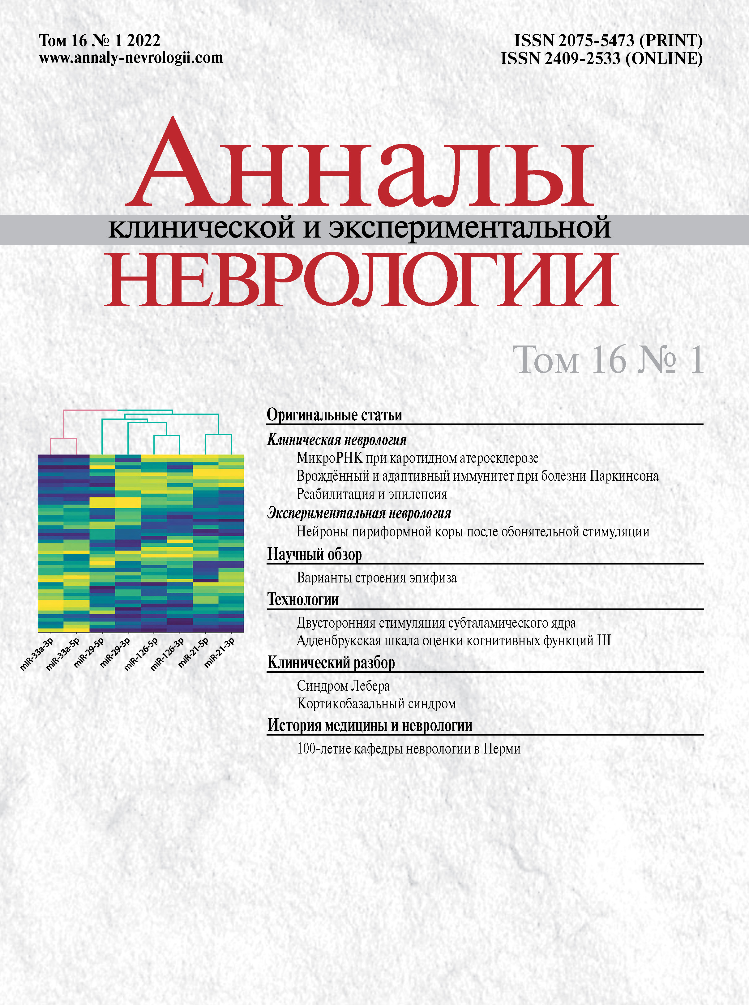
Materials and methods. The study included 50 people (median age 66 [61; 71] years, 58% men) with cerebrovascular disease secondary to CA. The patients were divided into two groups: 16 patients (32%) had ≥70% internal carotid artery (ICA) stenosis (main group), while the remaining 34 patients had <70% stenosis and formed the comparison group. Expression of the following microRNAs was measured: miR-126-5p, miR-126-3p, miR-29-5p, miR-29-3p, miR-33a-5p, miR-33a-3p, miR-21-5p and miR-21-3p.
Results. Compared to the comparison group, patients with a high degree of CA had reduced expression of miR-126-5p/-3p (4.8 and 5.9 vs. 8.5 and 7.6, respectively; p < 0.001) and miR-29-3p (7.6 vs. 10.3; p < 0.001), while miR-33a-5p expression was elevated (46.3 vs. 40.0; p < 0.05). Cluster analysis confirmed typical expression patterns of these microRNAs in patients with varying degrees of ICA stenosis. Significant negative correlations were also found between the degree of stenosis and expression of miR-126-5p (ρ = –0.83; р < 0.05), miR-126-3p (ρ = –0.64; р < 0.05) and miR-29-3p (ρ = –0.62; р < 0.05).
Conclusion. Based on an analysis of patients with cerebral atherosclerosis, the studied microRNAs can be divided into proatherogenic (miR-33a-5p/-3p) and atheroprotective (miR-126-5p/-3p, miR-29-3p, and mir-21-5p/-3p). These biomarkers can be diagnostically useful in predicting the risk of both CA progression and acute cerebrovascular accidents, yet prospective studies are required.
 5-13
5-13


Features of innate and adaptive immunity in patients with Parkinson's disease
Abstract
Introduction. T cells play a significant role in neuroinflammation in Parkinson's disease (PD). Gamma delta T cells are an under-researched 'minor' subpopulation of T cells. An assessment of the immune system in patients with PD, with a focus on γδТ cells, provides new data on the pathogenesis of neurodegenerative diseases.
The aim of the study was to examine the lymphocyte subpopulations, nonclassical γδТ cells, as well as cytokine production in patients with 3 stage PD complicated by motor fluctuations.
Materials and methods. We examined 20 patients with 3 stage PD receiving dopaminergic combination therapy (main group) and 20 age-matched patients with chronic cerebrovascular disease (comparison group). Considering the suspected role of chronic constipation in maintaining dysbiosis and chronic inflammation in patients with PD, the presence of constipation was an inclusion criterion for this study. The subpopulation profile of the peripheral blood lymphocytes was assessed using flow cytofluorometry, as well as cytokine levels using enzyme linked immunosorbent assay.
Results. It was found that the number of mature CD3+ T cells with αβ or γδ chains as the T-cell receptors (TCR) in the lymphocyte population was significantly lower in patients with PD — median 74% (57.3–83.5)) than in the comparison group (median 80% (73.0–86.0); р = 0.014. There was also a statistically significant reduction in the number of CD3+CD56+ natural killer (NK) T cells in the group of patients with PD vs. the comparison group — 4.7% (1.3–7.7) vs. 7.8% (0.8–24); р = 0.036. At the same time, the number of CD3–CD56+ NK cells was significantly higher in the group of patients with PD (16.4% (9–34)) vs. the comparison group — 8.7% (5–15); р = 0.001. Moreover, the main group had a statistically significantly higher number of activated CD3–CD8+ NK cells — 7% (4.5–13.5) vs. the comparison group — 3.5% (0.86–4.9); р < 0.001. Out of the total number of γδТ cells, the TCRγδ CD4+CD8– subpopulation was statistically smaller in the group of patients with PD — 13.6% (6.2–27.0) than in the comparison group — 29.8% (4.0–52.1); р = 0.016. The study of cytokine levels in the group of patients with PD showed a significant increase in the induced production of interleukin-1β (IL-1β), as well as a high (aberrant) spontaneous production of IL-10, which was 227.5 pg/ml in patients with PD when the normal range is 0–23 pg/ml. The correlation analysis showed that the TCRγδ CD4+CD8– subpopulation and cytokines in the group of patients with PD had a statistically significant (p = 0.048) negative correlation with the induced production of IL-10 (r = –0.745) and a significant (p = 0.042) positive correlation with the induced production of the pro-inflammatory cytokine IL-1β (r = 0.648). There was a trend towards increased spontaneous production of IL-10 (r = –0.602; p = 0.0506) as the level of the TCRγδ CD4+CD8– T helper cells decreased.
Conclusion. Changes were found in the blood of patients with PD, which indicate a chronic inflammatory process: increased number of CD3–CD56+ NK cells, including activated CD3–CD8+ cells, and increased production of pro-inflammatory cytokine IL-1β and anti-inflammatory cytokine IL-10. A decrease was found in the level of a minor subpopulation of γδT cells, TCRγδ CD4+CD8–. The correlation found between this subpopulation and the production of pro- and anti-inflammatory cytokines indicates its role in regulation of chronic inflammation in PD.
 14-23
14-23


Rehabilitation of young children with movement disorders and epilepsy: rational approach and efficacy
Abstract
Introduction. Epilepsy is one of the most common chronic nervous system disorders. Epilepsy in a child requiring physical, psychological and speech therapy significantly reduces its scope and decreases the likelihood of recovery.
The aim of the study was to assess the efficacy and safety of a rehabilitation programme for young children with movement disorders and concomitant epilepsy.
Materials and methods. Simple randomization was used to divide 123 children aged 9–24 months into four groups: three main groups and one comparison group. Patients in group 1 received traditional massage, excluding the cervical region, as their rehabilitation. Patients in group 2 received kinesiotherapy (Vojta therapy) in addition to traditional massage. Children in group 3 participated in a comprehensive programme, including traditional massage and kinesiotherapy (Vojta therapy). Children in the control group did not receive rehabilitation.
Results. A statistically significant improvement in the psychomotor development parameters was observed after a course of medical rehabilitation. It was more significant when the epileptic focus was localized in the right hemisphere or the patient had generalized epilepsy. The outcome was less favourable in multifocal epilepsy and when the epileptic focus was present on the convex surface of the left hemisphere. The third group noted a statistically significant improvement in the GMFCS scores by the end of the comprehensive rehabilitation course. There were no epileptic seizures seen on repeat EEG recordings during the medical rehabilitation and one month after its completion.
Conclusion. A comprehensive approach to planning a course of rehabilitation ensures its efficacy. The location of the epileptic focus and the distribution of epileptic activity along the convex surface of the brain determines the outcome of medical rehabilitation. An increased epileptiform activity index on EEG without signs of clinical deterioration requires more careful patient monitoring but, nevertheless, is not a reason to completely cancel rehabilitation measures.
 24-31
24-31


Expression of GABAergic and glutamatergic neurons after olfactory stimulation in the mouse piriform cortex during postnatal development
Abstract
Introduction. The control of the survival and differentiation of immature neurons in the piriform cortex of rodents, which can transform into GABAergic and/or glutamatergic neurons under the influence of olfactory stimuli, is an important factor for prevention of neurological dysfunction.
The aim of the study was to assess the expression of GABAergic and glutamatergic neurons after olfactory stimulation (OS) in the mouse piriform cortex during postnatal development.
Materials and methods. The study was carried out on CD1 male mice aged 2 (n = 20; group Р2), 21 (n = 20; group Р21) and 60 (n = 20; group Р60) days. The mice were presented with olfactory stimuli, and brain tissue was collected for immunohistochemical analysis 2 hours, 24 hours and 7 days later, to assess glutamic acid decarboxylase 67 (GAD67) and vesicular glutamate transporter 1 (VGlut1) expression.
Results. OS in the group P2 animals increased VGlut1 expression in the first 2 hours after OS, followed by a return to baseline level by day 7, while GAD67 expression showed no significant changes. The animals in group P21 showed increased expression of VGlut1 and GAD67 two hours after OS, followed by a significant decrease. Expression of both molecules demonstrated a statistically significant increase in the group P60 animals 24 hours after OS, and remained at the same level on day 7 (GAD67) or returned to baseline levels (VGlut1).
Conclusion. OS increases the number of GABAergic (GAD67+) и glutamatergic (VGlut1+) neurons in the piriform cortex (P60). The predominance of glutamatergic effects is a possible mechanism for associative memory cell recruitment.
 32-38
32-38


Reviews
Pineal gland: structural variants and their role in neurological and psychiatric disorders
Abstract
The pineal gland is a small and poorly studied neuroendocrine gland located in the epithalamus. There is growing interest in the pineal gland due to its role in regulating human biological rhythms, which is associated with melatonin production, and its close neuroendocrine link between the brain's hormonal and neurally mediated activity. The paper examines the anatomical and physiological features of the pineal gland, its structural variations, and the role of the melatonin it produces in the pathogenesis of several mental and neurological disorders.
 39-45
39-45


Technologies
Bilateral stimulation of the subthalamic nucleus under local and general anaesthesia
Abstract
Introduction. Bilateral stimulation of the subthalamic nucleus (STN) is successfully used to treat advanced stages of Parkinson's disease. The standard surgical technique includes microelectrode recording and intraoperative stimulation. The introduction of 3T MRI into clinical practice and new impulse sequences have led to the question of whether the surgery can be performed under general anaesthesia.
Aim of the study: to compare the efficacy and safety of bilateral stimulation of STN in patients with Parkinson's disease, using 3T MRI under local and general anaesthesia.
Materials and methods. This prospective, randomized controlled study included 40 patients, who underwent electrode implantation using 3T MRI. The patients in the main group (n = 20) had electrodes implanted under general anaesthesia, while the control group (n = 20) had local anaesthesia, intraoperative stimulation and microelectrode recording. The patients’ motor status, quality of life, cognitive function and required levodopa dose were evaluated after 6 months.
Results. The results of 6 months of stimulation were obtained from 30 patients (15 from the main group and 15 from the control group). Reduced motor impairment was observed in both groups as measured on the UPDRS III scale during the 'off' time, with a reduction of 68.7% in the main group and 74.7% in the control group. Improved quality of life, reduced drug-induced motor complications and a reduction in the levodopa dose were also comparable in both groups. No intraoperative haemorrhagic complications were recorded during the study.
Conclusions. Preliminary analysis of the data showed that with high-quality visualization of the STN, electrode implantation under anaesthesia is equally effective to awake surgery.
 46-52
46-52


The Addenbrooke’s Cognitive Examination III (ACE-III): linguistic and cultural adaptation into Russian
Abstract
Introduction. Timely identification of cognitive impairment is very important, with standardized screening instruments required to assess the cognitive status. However, the arsenal of such screening scales available to clinicians in Russia is limited and requires expansion. According to numerous international studies, the Addenbrooke’s Cognitive Examination III (ACE-III) has the necessary sensitivity and specificity, which speaks well for developing and validating a Russian language version.
The aim of the study was the linguistic and cultural adaptation of the Addenbrooke’s Cognitive Examination III (ACE-III).
Materials and methods. A forward and back translation was performed of three versions of the scale and the scoring guidelines. A preliminary version of the ACE-III was developed, pilot testing of the preliminary version was conducted, and a final Russian language version was then developed with the help of a philologist/linguist, and experts in neuropsychology and neurology, who work specifically with patients with cognitive impairments. Pilot testing of the preliminary version of the ACE-III involved 16 neurological patients at the Research Center of Neurology and the Pirogov National Medical and Surgical Centre, who were aged 37–74 (60.25 ± 10.8) years and 56% of whom were women. The patients’ clinical condition corresponded to the diagnostic criteria for cerebrovascular disease (n = 12), Parkinson's disease (n = 3) and spinocerebellar ataxia (n = 1).
Results. Neither the subjects nor the examiners had any difficulty in understanding the instructions or the content during testing. Further work was done based on the results of the pilot testing, and three final versions of the scale (A, B and C) were accepted, as well as the scoring guidelines, a link to which is provided in the article.
Conclusion. The obtained results indicate that the developed version of the ACE-III can be understood by the Russian-speaking population and can be used in clinical practice. At the time of article publication, research is being conducted to assess the psychometric properties of the final Russian language version.
 53-58
53-58


Clinical analysis
Clinical observations of Leber hereditary optic neuropathy with and without neurological symptoms
Abstract
Three case studies of adults with Leber hereditary optic neuropathy with and without neurological symptoms are presented. An elevated blood lactate level and changes in the activity of the mitochondrial enzymes in peripheral blood lymphocytes were noted. Mutations in the mitochondrial DNA (G3460A in one patient and G11778A in two patients) were found. The patient with a G3460A mutation, in addition to reduced visual acuity, was diagnosed with a cerebellar disorder due to cerebellar vermis hypoplasia. These changes are indications for prescribing energotropic drugs (idebenone, carnitine); carnosine can also be prescribed. These case studies show that patients with optic nerve atrophy should be assessed for Leber hereditary optic neuropathy. Differential diagnosis with multiple sclerosis should be performed, since this condition often presents as optic neuropathy.
 59-63
59-63


Corticobasal syndrome as a phenotype of various neurodegenerative disorders: a case series
Abstract
Corticobasal syndrome (CBS) is a variant of atypical parkinsonism. The underlying cause may be corticobasal degeneration or other proteinopathies, which can be verified only after studying specific biomarkers. The disease aetiology in CBS needs to be established to determine the disease prognosis. It can also affect the choice of pathogenetic treatment due to the differences in the molecular pathogenesis of proteinopathies that cause neurodegenerative processes. Four clinical cases of CBS are presented: in patients with four-repeat tauopathy, Alzheimer's disease, frontotemporal dementia and Creutzfeldt–Jakob disease. Examples are provided of the clinical, genetic and biochemical biomarkers available for differential diagnosis of CBS.
 64-70
64-70


Historical articles
For the 100th anniversary of the Department of neurology in Perm
Abstract
October 2021 marks the 100th anniversary of the founding of the Department of neurology and medical genetics at the Perm State Medical University. The article presents a historical perspective, the main clinical and research achievements, and development prospects for the department.
 71-75
71-75


